Periapical X-Rays

What Are Periapical X-Rays?
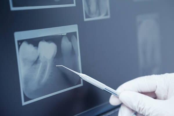
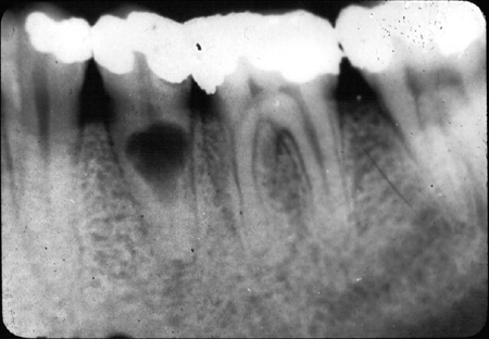
Periapical X-rays are an essential diagnostic tool in dental care. Unlike panoramic or bitewing X-rays, which capture broader views of the oral cavity, periapical X-rays focus on a specific area of the mouth. They provide detailed imaging of the entire tooth—showing everything from the crown (visible part above the gumline) to the root (embedded in the jawbone)—as well as the surrounding bone structure.
These X-rays are vital for diagnosing and treating dental conditions that may not be visible during a routine exam. They help identify issues such as:
- Deep tooth decay
- Abscesses or infections at the tooth root
- Bone loss around teeth
- Fractures or damage to teeth
- Cysts, tumors, or other abnormalities
Periapical X-rays allow dentists to develop accurate treatment plans, ensuring optimal care for patients. Before deciding on whether Periapical X-Rays are right for you, there are some things you should know:
- Who Needs Periapical X-Rays?
- Benefits Of Periapical X-Rays
- How Much Do Periapical X-Rays Cost?
- Steps In The Periapical X-Rays Procedure
- Frequently Asked Questions About Periapical X-Rays
If you have any further questions about Periapical X-Rays or other dental services offered at Atlas Dental, please contact us.

Free phone consultation
Have questions about Periapical X-Rays? Schedule a free phone consultation with our Toronto dentist.

5 star google reviews
Our patients love us! See for yourself why more and more people are choosing Atlas Dental for their Periapical X-Rays.

Book Periapical X-Rays Scan Online
We provide quick and convenient Periapical X-Rays Scan Service in Toronto. Book Online.
Who Needs Periapical X-Rays?
Dentists may recommend periapical X-rays in the following scenarios:
- Tooth Pain or Sensitivity: Identifying the source of unexplained discomfort, such as deep cavities or root infections.
- Gum Disease: Detecting bone loss or damage caused by periodontal disease for effective treatment.
- Dental Trauma: Assessing fractures or dislodged teeth caused by injury.
- Root Canal Therapy: Monitoring treatment progress and ensuring the root canal is thoroughly treated.
- Dental Implants: Evaluating jawbone health and planning the precise placement of implants.
- Monitoring Chronic Conditions: Tracking changes in dental cysts, tumors, or impacted teeth over time.
By using periapical X-rays, dentists can gather vital information to diagnose and treat various dental issues effectively, ensuring you maintain a healthy and pain-free smile. If you have further questions about Periapical X-Rays, please contact us.

Benefits Of Periapical X-Rays
Periapical X-rays offer numerous benefits, including:
- Detailed Imaging: A clear view of the entire tooth and surrounding bone structure.
- Accurate Diagnoses: Early detection of dental issues like decay, infections, and fractures.
- Effective Treatment Planning: Information critical for successful procedures such as root canals, extractions, or implant placement.
- Monitoring Progress: Ensuring ongoing treatments like gum disease therapy are effective.
- Detection of Hidden Problems: Uncovering abnormalities like cysts or impacted teeth before symptoms arise.
- Low Radiation Exposure: Modern digital X-rays use minimal radiation, ensuring patient safety.
Overall, the advantages of periapical X-rays make them an essential part of maintaining oral health. They provide critical insights that help in the early detection, accurate diagnosis, and effective treatment of various dental conditions. If you have further questions about Periapical X-Rays, please contact us.
Cost of Digital Dental X-Rays Images
The cost of Digital Dental X-Ray Images will depend on the type of image taken as well as the number of images. Example x-ray codes in the Ontario Dental Association’s Suggested Fee Guide appear as follows:
- 02111 – Single image: $41
- 02112 – Two images: $48
- 02113 – Three images: $56
- 02114 – Four images: $63
- 02115 – Five images: $74
- 02116 – Six images: $82
- 02117 – Seven images: $92
- 02141 – Single image: $41
- 02142 – Two images: $48
- 02143 – Three images: $56
- 02144 – Four images: $61
Radiographs, Regional/Localized
- 02101 – Radiographs, Complete Series (minimum of 12 images incl. bitewings): $166
- 02102 – Radiographs, Complete Series (minimum 16 images, incl. bitewings): $179
- 02601 – Single images: $84
Dental X-Rays are considered a basic service under all dental insurance plans and should be covered to your maximum insurable limit, but be sure to find out from your dental insurance plan provider how much you are eligible for before going ahead with dental treatment. Our fees are consistent with the ODA Fee Guide.
For patients without dental insurance, Atlas Dental is pleased to offer dental financing through iFinance Dentalcard. Affordable payment plans start at 7.95% for terms of 6 months to 6 years. To learn more about Dentalcard dental treatment financing, follow this link.
Steps In The Periapical X-Ray Procedure
Here’s a step-by-step overview of the periapical X-ray process:
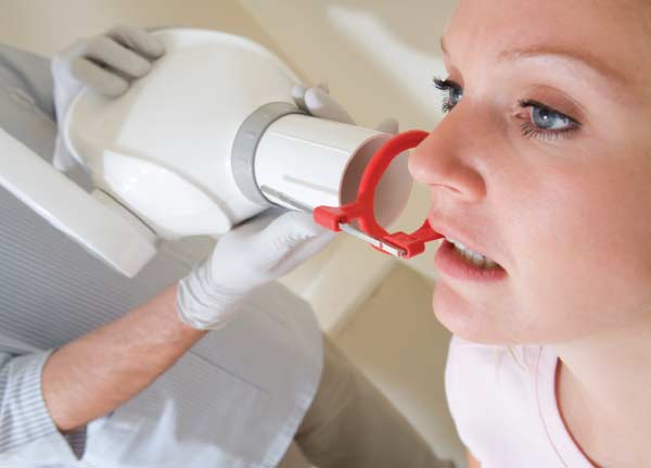
- Preparation: You’ll wear a lead apron to protect against radiation exposure, and the dental technician will position the X-ray sensor or film in your mouth.
- Positioning: The technician will guide you to sit or stand in the correct position and place a bite block for stabilization.
- Image Capture: The X-ray machine is aligned to target the specific tooth or area. You’ll need to remain still for a few seconds while the image is taken.
- Image Analysis: The dentist will review the X-ray immediately, discussing findings and recommending any necessary treatments.
By following these steps, dentists can obtain clear and accurate periapical X-ray images to aid in diagnosis and treatment planning for their patients’ dental care needs. If you have further questions about Periapical X-Rays, please contact us.

Frequently Asked Questions About Periapical X-Rays
- Are Periapical X-rays safe?
Yes, modern periapical X-rays use minimal radiation, and precautions such as lead aprons are used to protect patients.
- Can I have a periapical X-ray if I’m pregnant?
Inform your dentist if you’re pregnant. While X-rays are generally avoided during pregnancy, they may still be performed in emergencies with added precautions.
- How long does a periapical X-ray take?
The process is quick, typically taking less than five minutes per image.
- Are periapical X-rays painful?
No, the procedure is painless. You may feel slight pressure from the sensor or film in your mouth, but it’s not uncomfortable.
At Atlas Dental, we prioritize patient care and comfort. With state-of-the-art equipment and experienced dental professionals, we ensure your X-rays are accurate and safe. Contact us today to book your periapical X-ray and take the first step toward a healthier smile!

